Lucia Hong, Elaine Yu
A 70-year-old male with a history of cirrhosis, COPD, HTN, T2DM, and large abdominal wall hernia who presented after being found down in his home by a neighbor. Upon arrival, the patient was hypotensive with systolic blood pressures in the 70s and in atrial fibrillation with RVR with heart rates in the 170s. He received 500mL intravenous fluids prior to arrival and was transported on supplemental oxygen. The patient was altered and unable to provide history.
On physical examination, the patient appeared acutely ill and minimally responsive. Mucous membranes were dry. Cardiovascular examination demonstrated tachycardia with an irregular rhythm. Lung examination revealed bilateral breath sounds without focal wheezes or stridor. The abdomen was distended with generalized tenderness and a large non-reducible abdominal wall hernia. Extremities were warm and perfused without significant peripheral edema. Neurologic examination demonstrated altered mental status with intermittent command following and spontaneous movement of all extremities.
Synchronized cardioversion was performed following sedation with fentanyl and midazolam, which resulted in sinus tachycardia with improvement in heart rate and blood pressure.
Vital Signs: BP: 94/62 | HR: 102 | RR: 25 | Temp: 100 °F| SpO₂: 99% on 5L O2
Following cardioversion, RUSH was performed with findings of a small pericardial effusion, a plethoric inferior vena cava with minimal respiratory variation, and abnormal right ventricular wall motion with apparent right ventricular enlargement (Figure 1).
Additionally, intra-abdominal free fluid concerning for ascites was also seen (Figure 2). These findings prompted further evaluation of cardiogenic, obstructive, and distributive shock.


Labs: WBC 13.3, Hgb 6.9, lactate 2.1, troponin 192 -> 189, D-dimer 26,069
Imaging
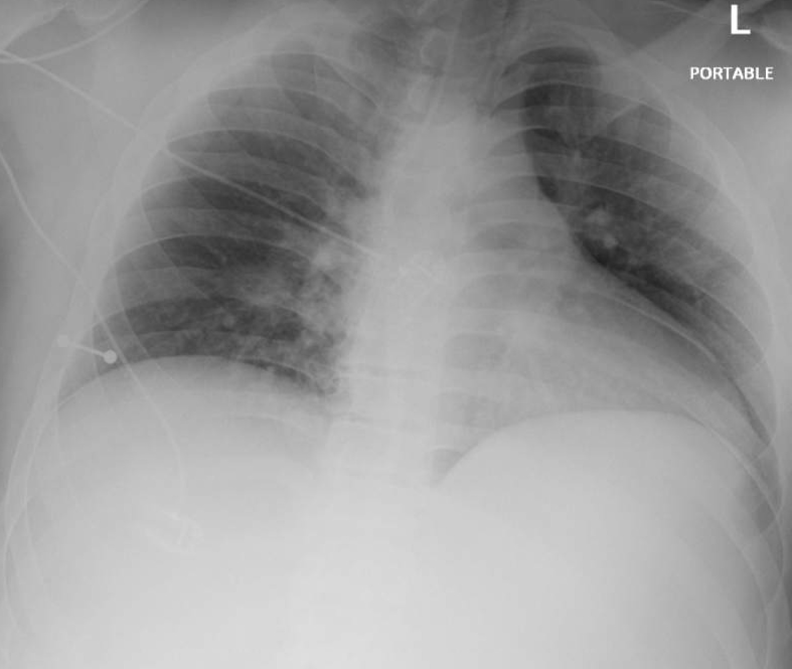
CTA PE: No definite pulmonary embolism. Mild volume overload, probably cardiogenic.
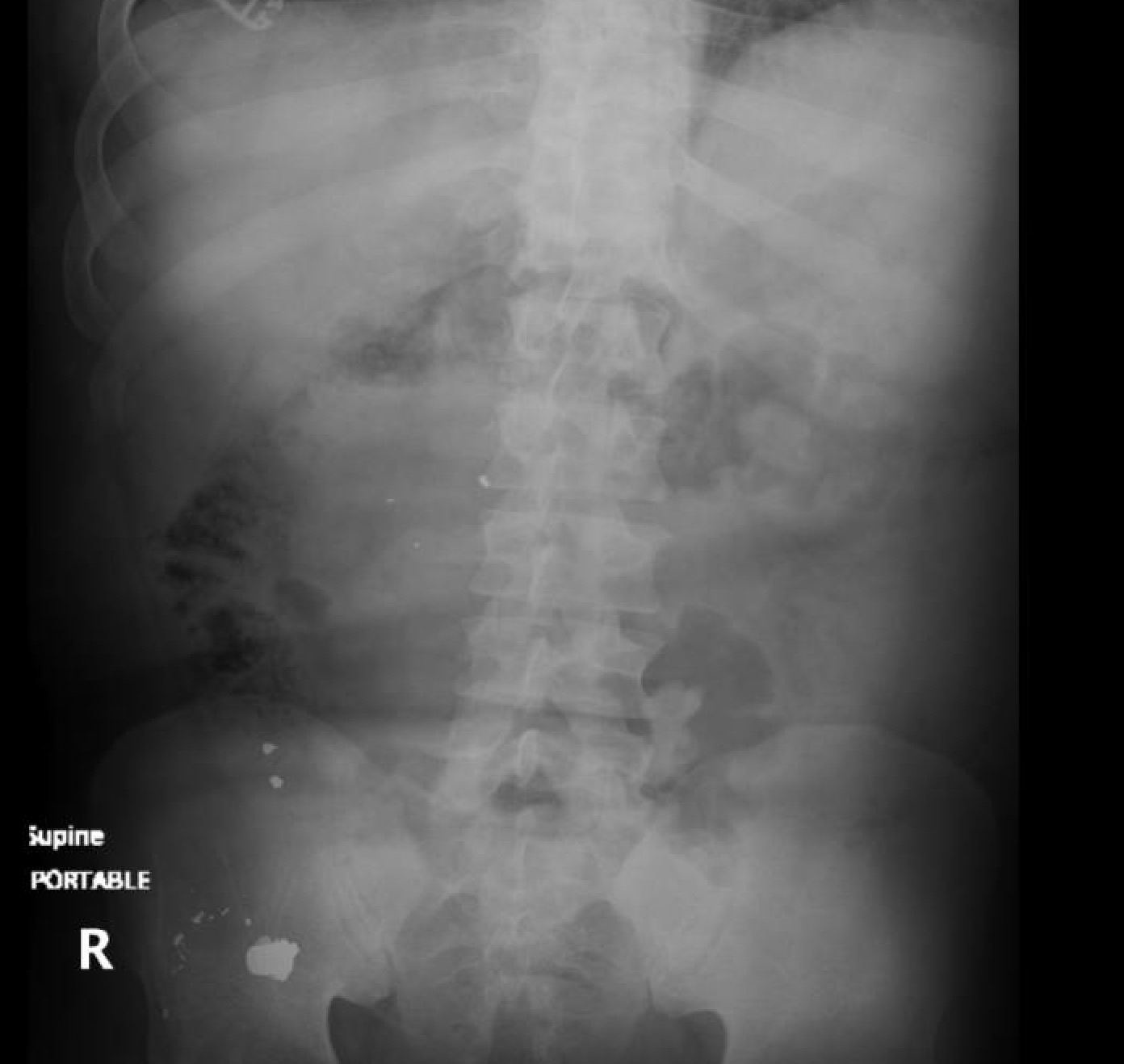
CT Abdomen/Pelvis with contrast: 4.7 cm left anterior bladder wall abscess. Large volume ascites.
Discussion
Undifferentiated hypotension in the emergency department presents a diagnostic challenge, particularly in patients with multiple comorbidities and competing etiologies of shock. Rapid Ultrasound in Shock and Hypotension (RUSH) examination has emerged as a critical bedside tool allowing evaluation of physiologic contributors to shock prior to definitive diagnostic testing. The RUSH protocol integrates focused cardiac, vascular, pulmonary, and abdominal ultrasound assessment to assess hypovolemic, distributive, cardiogenic, or obstructive etiologies.3 Incorporation of early bedside ultrasound has been shown to alter the presumed category of shock in up to 50% of patients presenting with nontraumatic hypotension.4 RUSH is associated with faster diagnostic clarification and earlier targeted therapy in critically ill emergency department patients.5 The utilization of POCUS has demonstrated high specificity for detecting right ventricular strain patterns associated with obstructive shock states.6 Furthermore, POCUS can improve evaluation of volume status and reduce potentially harmful fluid overload in critically ill patients.7
In this case, the patient presented with hypotension, altered mental status, and atrial fibrillation with RVR. Multiple or mixed shock etiologies were plausible, including septic shock from intra-abdominal infection, cardiogenic shock related to arrhythmia or myocardial injury, and obstructive shock with pulmonary embolism. Additionally, hypovolemia was also considered given an initial Hgb 6.9. Identification of right ventricular wall abnormalities increased clinical suspicion for obstructive pathology, and a subsequent D-dimer was noted to be significantly elevated. CTA PE was completed that ruled out pulmonary embolism and demonstrated volume overload from a likely cardiogenic cause. Further CT images identified a bladder abscess as a source of sepsis. Additionally, RUSH examination findings contributed to cautious fluid administration and prompted consideration of alternative shock mechanisms.
This case highlights how POCUS guides subsequent decision-making. As emphasized in current American College of Emergency Physicians guidelines, POCUS serves as an extension of the physical examination and plays an increasingly central role in the early evaluation of critically ill patients in the emergency department.8
References
1. Kansara T, Quesada F, Park H, Ghosh K, Saeed M. McConnell’s Sign Still Holds Its Value: A Lesson Learned From Two Cases. Cureus. 2019;11(11):e6240. doi:10.7759/cureus.6240
2. Zuidewind P. Cirrhosis and portal hypertension. Case study, Radiopaedia.org. Published June 21, 2020. https://radiopaedia.org/cases/cirrhosis-and-portal-hypertension-1
3. Perera P, Mailhot T, Riley D, Mandavia D. The RUSH exam: Rapid ultrasound in shock in the evaluation of the critically ill. Emerg Med Clin North Am. 2010;28(1):29–56.
4. Jones AE, Tayal VS, Sullivan DM, Kline JA. Randomized, controlled trial of immediate versus delayed goal-directed ultrasound to identify the cause of nontraumatic hypotension in emergency department patients. Crit Care Med. 2004;32(8):1703–1708.
5. Atkinson PRT, Milne J, Diegelmann L, et al. Does point-of-care ultrasonography improve clinical outcomes in emergency department patients with undifferentiated hypotension? A systematic review and meta-analysis. Resuscitation. 2018;127:1–9.
6. Nazerian P, Vanni S, Volpicelli G, et al. Accuracy of point-of-care multiorgan ultrasonography for the diagnosis of pulmonary embolism. Chest. 2014;145(5):950–957.
7. Marik PE, Monnet X, Teboul JL. Hemodynamic Parameters to Guide Fluid Therapy. Ann Intensive Care. 2011;1:1.
8. American College of Emergency Physicians. Emergency Ultrasound Guidelines. Ann Emerg Med. 2017;69(5):e27–e54.


")


